Show all abstracts Show selected abstracts Add to my list |
|
EDITORIALS |
|
|
|
Staging in giant vestibular Schwannoma surgery |
p. 209 |
Isabel Cuervo-Arango Herreros, Alvaro Campero DOI:10.4103/0976-3147.133549 PMID:25002756 |
[HTML Full text] [PDF] [Mobile Full text] [EPub] [PubMed] [Sword Plugin for Repository]Beta |
|
|
|
|
|
|
Microsurgical training model for residents to approach the orbit and the optic nerve in fresh cadaveric sheep cranium |
p. 210 |
Thomas Mucke DOI:10.4103/0976-3147.133551 PMID:25002757 |
[HTML Full text] [PDF] [Mobile Full text] [EPub] [PubMed] [Sword Plugin for Repository]Beta |
|
|
|
|
|
|
ORIGINAL ARTICLES |
 |
|
|
 |
The effect of craniectomy size on mortality, outcome, and complications after decompressive craniectomy at a rural trauma center  |
p. 212 |
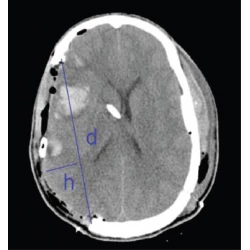
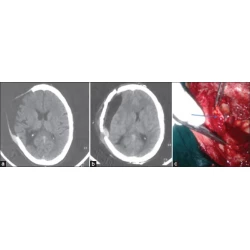
Cara L. Sedney, Terrence Julien, Jacinto Manon, Alison Wilson DOI:10.4103/0976-3147.133555 PMID:25002758Introduction: Decompressive craniectomy (DC) has increasing support with current studies suggesting an improvement in both survival rates and outcomes with this intervention. However, questions surround this procedure; specifically, no evidence has indicated the optimal craniectomy size. Larger craniectomy is thought to better decrease intracranial pressure, but with a possible increase in complication rates. Our hypothesis is that a larger craniectomy may improve mortality and outcome, but may increase complication rates. Materials and Methods: A retrospective observational therapeutic study was undertaken to determine if craniectomy size is related to complication rates, mortality, or outcome. Our institution's Trauma Registry was searched for patients undergoing DC. Craniectomy size was measured by antero-posterior (AP) diameter. Mortality, outcome (through admission and discharge Glasgow Coma Score and Glasgow Outcome Scale), and complications (such as re-bleeding, re-operation, hygroma, hydrocephalus, infection, and syndrome of the trephined) were noted. Complications, mortality, and outcome were then compared to craniectomy size, to determine if any relation existed to support our hypothesis. Results: 20 patients met criteria for inclusion in this study. Craniectomy size as measured by AP diameter was correlated with a statistically significant improvement in mortality within the group. All patients with a craniectomy size less than 10 cm died. However, outcome was not significantly related to craniectomy size in the group. Similarly, complication rates did not differ significantly compared to craniectomy size. Discussion: This study provides Level 3 evidence that craniectomy size may be significantly related to improved mortality within our group, supporting our initial hypothesis; however, no significant improvement in outcome was seen. Similarly, in contrast to our hypothesis, complication rates did not significantly correlate with craniectomy size. |
[ABSTRACT] [HTML Full text] [PDF] [Mobile Full text] [EPub] [Citations (2) ] [PubMed] [Sword Plugin for Repository]Beta |
|
|
|
|
|
 |
Clinical profile and outcome of cerebral venous sinus thrombosis at tertiary care center  |
p. 218 |
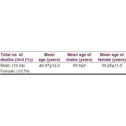
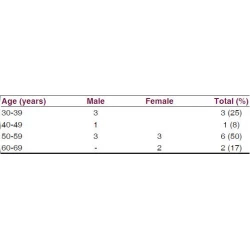
Virendra C. Patil, Kushal Choraria, Neeraj Desai, Sumit Agrawal DOI:10.4103/0976-3147.133559 PMID:25002759Background: Thrombosis of the cerebral venous sinuses (CVST) is an uncommon form of stroke, usually affecting young individuals. Clinical features of CVST are diverse, and for this reason, high degree of clinical suspect is mandatory to diagnose the conditions. Materials and Methods: This study was conducted over a period of 1 year (Jan 2011 to Dec 2011). This was a retrospective, observational, and noninterventional study. This study was conducted in the Department of Medicine at a tertiary care teaching center. Total 50 patients where diagnosis of CVST was confirmed by computed tomography/magnetic resonance imaging brain venogram were included in this study. All patients with diagnosis of CVST were treated according to the standard protocol and guidelines. Statistical Analysis: The mean and standard deviation were obtained. The Chi-square test was used to analyze the data and P < 0.05 was considered as statistically significant. Results: Of total 50 patients with diagnosis of CVST, 21 (42%) were males and 29 (58%) were females with 39 ± 10 years and 29 ± 7 years, respectively. Total 45 (90%) patients presented with symptoms of headache and vomiting, 13 (26%) had seizures, 12 (24%) had hemiplegia, and 19 (38%) had fever. A total of 13 (26%) patients had papilledema on fundoscopy. Total 9 (31%) out of 29 patients had diagnosis of CVST during peripartum period. Total 12 (24%) patients had hyperhomocysteinemia. Total 23 (46%) patients had sagittal sinus thrombosis, 10 (20%) had multiple sinus thrombosis, 16 (32%) had sigmoid/transverse sinus thrombosis. There was 1 (2%) patient who had bilateral cavernous sinus thrombosis, who presented with bilateral proptosis, conjunctival congestion, and external ophthalmoplegia with a history of acute or chronic maxillary and sphenoid sinusitis. Total 38 patients had evidence of infection in the form of fever, paranasal sinus (PNS) infections, Chronic suppurative otitis media (CSOM). Total 19 (38%) patients had a history and evidence of dehydration. Total 8 (16%) patients died during the course of treatment and 42 (84%) were discharged with partial and/or total recovery. Three (6%) patients required neurosurgical intervention in the form of decompressive craniotomy. Eight (16%) patients died with cerebral edema with transtentorial herniation. The mean age of death in male was significantly greater than in female patients with P < 0.02. Majority of patients succumbed had sigmoid, transverse, and/or multiple sinus involvement. Patients with multiple sinus thrombosis had greater case fatality rate. Conclusions: The current study highlights the burden of CVST in the study population with headache and vomiting, which was the most common presenting complaint. The superior sagittal sinus thrombosis was the most common and bilateral cavernous sinus thrombosis was the uncommon affection in CVST. One third of female population was affected in peripartum period. The infection and/or dehydration was the most commonly associated precipitating event for development of CVST and more than one fifth of the population had evidence of hyperhomocysteinemia. Mortality was more in patients with affection of sigmoid, transverse, and/or multiple sinus involvement in male patients and superior sagittal sinus thrombosis in female patients. The treatment of CVST has to be aggressive as morbidity and mortality is relatively minimal compared with the arterial stroke. |
[ABSTRACT] [HTML Full text] [PDF] [Mobile Full text] [EPub] [Citations (2) ] [PubMed] [Sword Plugin for Repository]Beta |
|
|
|
|
|
 |
Staging in giant vestibular schwannoma surgery: A two consecutive day technique for complete resection in basic neurosurgical setups |
p. 225 |
Deepak Bandlish, Nilay Biswas, Sumit Deb DOI:10.4103/0976-3147.133560 PMID:25002760Introduction: Vestibular schwannomas constitute 8% of all intracranial tumors. A majority of vestibular schwannomas are sporadic and unilateral. Giant vestibular schwannomas are seen in our country due to the late diagnosis and long duration of symptoms before diagnosis. These giant schwannomas are challenging to manage as most of the patients are having brainstem compression. Materials and Methods: Twelve cases of a giant vestibular schwannoma were operated in our department between May 2011 and December 2012. Vestibular schwannomas with a maximal diameter of more than 4 cm were defined as a giant vestibular schwannoma. All the patients had a unilateral vestibular schwannoma. Performance status of all the patients were graded as per the Karnofsky performance score. Pre-operative assessment of 5 th , 7 th , 8 th and lower cranial nerve status was done in all cases. Ventriculoperitoneal shunting was done pre-operatively in all cases. All patients were operated through retromastoid suboccipital craniectomy and retrosigmoid approach. These patients were operated in two stages in two consecutive days with overnight elective ventilation in ICU. Ultrasonic aspirator and nerve monitoring techniques were not used. Results: Giant acoustic schwannomas can be safely resected completely by a staged resection on two consecutive days without any increased morbidity or mortality. This technique may be employed to achieve complete resection of such lesions without deterioration of facial nerve function in institutions which do not have advanced facilities like nerve monitoring or ultrasonic aspirator. |
[ABSTRACT] [HTML Full text] [PDF] [Mobile Full text] [EPub] [PubMed] [Sword Plugin for Repository]Beta |
|
|
|
|
|
 |
Intracranial nonvestibular neurinomas: Young neurosurgeons' experience  |
p. 231 |
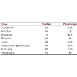
Forhad Hossain Chowdhury, Mohammod R. Haque, Khandkar A. Kawsar, Mainul H. Sarker, Mahmudul Hasan, Atul H. Goel DOI:10.4103/0976-3147.133566 PMID:25002761Background and Objectives: Neurinoma arising from other than nonvestibular cranial nerves is less prevalent. Here we present our experiences regarding the clinical profile, investigations, microneurosurgical management, and the outcome of nonvestibular cranial nerve neurinomas. Materials and Methods: From January 2005 to December 2011, the recorded documents of operated nonvestibular intracranial neurinomas were retrospectively studied for clinical profile, investigations, microneurosurgical management, complications, follow-up, and outcomes. Results: The average follow-up was 24.5 months. Total number of cases was 30, with age ranging from 9 to 60 years. Sixteen cases were males and 14 were females. Nonvestibular cranial nerve schwannomas most commonly originated from trigeminal nerve followed by glossopharyngeal+/vagus nerve. There were three abducent nerve schwannomas that are very rare. There was no trochlear nerve schwannoma. Two glossopharyngeal+/vagus nerve schwannomas extended into the neck through jugular foramen and one extended into the upper cervical spinal canal. Involved nerve dysfunction was a common clinical feature except in trigeminal neurinomas where facial pain was a common feature. Aiming for no new neurodeficit, total resection of the tumor was done in 24 cases, and near-total resection or gross total resection or subtotal resection was done in 6 cases. Preoperative symptoms improved or disappeared in 25 cases. New persistent deficit occurred in 3 cases. Two patients died postoperatively. There was no recurrence of tumor till the last follow-up. Conclusion: Nonvestibular schwannomas are far less common, but curable benign lesions. Surgical approach to the skull base and craniovertebral junction is a often complex and lengthy procedure associated with chances of significant morbidity. But early diagnosis, proper investigations, and evaluation, along with appropriate decision making and surgical planning with microsurgical techniques are the essential factors that can result in optimum outcome. |
[ABSTRACT] [HTML Full text] [PDF] [Mobile Full text] [EPub] [PubMed] [Sword Plugin for Repository]Beta |
|
|
|
|
|
 |
Histological-subtypes and anatomical location correlated in meningeal brain tumors (meningiomas) |
p. 244 |
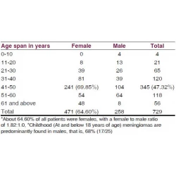
Abdul Rashid Bhat, Muhammed Afzal Wani, Altaf Rehman Kirmani, Altaf Umar Ramzan DOI:10.4103/0976-3147.133568 PMID:25002762Context: Not enough literature is available to suggest a link between the histological subtypes of intracranial meningeal brain tumors, called 'meningiomas' and their location of origin. Aim: The evidence of correlation between the anatomical location of the intracranial meningiomas and the histopathological grades will facilitate specific diagnosis and accurate treatment. Materials and Methods: The retrospective study was conducted in a single high-patient-inflow Neurosurgical Center, under a standard and uniform medical protocol, over a period of 30 years from December 1982 to December 2012. The records of all the operated 729 meningiomas were analyzed from the patient files in the Medical Records Department. The biodata, x-rays, angiography, computed tomography (CT) scans, imaging, histopathological reports, and mortality were evaluated and results drawn. Results: The uncommon histopathological types of meningiomas (16.88%) had common locations of origin in the sphenoid ridge, posterior parafalcine, jugular foramen, peritorcular and intraventricular regions, cerebellopontine angle, and tentorial and petroclival areas. The histopathological World Health Organization (WHO) Grade I (Benign Type) meningiomas were noted in 89.30%, WHO Grade II (Atypical Type) in 5.90%, and WHO Grade III (Malignant Type) in 4.80% of all meningiomas. Meningiomas of 64.60% were found in females, 47.32% were in the age group of 41-50 years, and 3.43% meningiomas were found in children. An overall mortality of 6.04% was noted. WHO Grade III (malignant meningiomas) carried a high mortality (25.71%) and the most common sites of meningiomas with high mortality were: The cerebellopontine angles, intraventricular region, sphenoid ridge, tuberculum sellae, and the posterior parafalcine areas. Conclusion: The correlation between the histological subtypes and the anatomical location of intracranial meningeal brain tumors, called meningiomas, is evident, but further research is required to establish the link. |
[ABSTRACT] [HTML Full text] [PDF] [Mobile Full text] [EPub] [Citations (2) ] [PubMed] [Sword Plugin for Repository]Beta |
|
|
|
|
|
 |
Clinical assessment of depression and type 2 diabetes in Morocco: Economical and social components |
p. 250 |
Salma Bensbaa, Loubna Agerd, Said Boujraf, Chadya Araab, Rachid Aalouane, Ismail Rammouz, Farida Ajdi DOI:10.4103/0976-3147.133576 PMID:25002763Background: The global prevalence of diabetes is increasing worldwide. In Morocco, diabetes and depression are major public health problems, requiring improvement in their care. Diabetes and depression are associated with morbidity and early mortality. This association contributes to raising the risk of the complications that occur, while causing higher suffering to patients, as also an increased cost toward healthcare. Aim: This study aims to assess the prevalence of depression in patients with type 2 diabetes (T2D), and identify the main risk factors for depression in this category of diabetic patients. Patients and Methods: Type 2 diabetic patients and older than 18 years of age were recruited. The exclusion criteria included being type 1 diabetic, pregnant woman, hospitalized patients, a history of neurological disorders, such as, stroke, infectious episidodes, and history of psychiatric disorders. The individual patient data was collected through individual and confidential interviews lasting 30 minutes, at the end of the diabetology consultation, by the same diabetologist, trained to use the psychometric scales that were needed. The Moroccan-Arabic version of the Beck diagnostic scale of depression was used. Patients assessed with depressive disorders were reviewed in a specialized psychiatric consultation. The statistical analysis was achieved by using SPSS package (version 17). We retained a threshold P value of 0.05. Settings and Design: A cross-sectional study was conducted that included adults with type 2 diabetes. The depression diagnosis was performed using the Arabic version of the Beck Depression scale. Statistical Analysis: We included 142 patients with type 2 diabetes, with an average age of 56.26 years. The prevalence of depression was 33.1%. The risk factors recognized for depression were, lack of social security, hypertension, and a history of type 2 diabetes of more than five years. Results and Conclusions: In this study, we have focused on the frequent association of 'Type 2 diabetes and depression' and the risk of mutual aggravation of both pathologies that might require multidisciplinary healthcare, as well as, improvement in the risk factors of depression through improved access to healthcare, with the extension of social security. The stability of the healthcare personnel involved in the treatment of both chronic diseases, including diabetes and hypertension screening, should also be considered for better management of psychiatric complications. |
[ABSTRACT] [HTML Full text] [PDF] [Mobile Full text] [EPub] [Citations (2) ] [PubMed] [Sword Plugin for Repository]Beta |
|
|
|
|
|
 |
Stent-assisted coiling of wide-necked intracranial aneurysms using the Solitaire AB stent |
p. 254 |
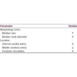
Vikram Huded, Rithesh R. Nair, Devashish D. Vyas, Bhumir N. Chauhan DOI:10.4103/0976-3147.133577 PMID:25002764Context: Stent-assisted coiling of wide-necked and complex intracranial aneurysms is an effective and feasible treatment option. The self-expanding, fully retrievable Solitaire AB (eV3, Irvine, CA, USA) stent is the latest neurovascular remodeling device available. To the best of our knowledge, there are no studies of Solitaire AB-assisted coiling of wide-necked intracranial aneurysms from India. Aim: Solitaire AB-assisted coiling of wide-necked intracranial aneurysms. Materials and Methods: The study was conducted in a tertiary care center with a dedicated Interventional Neurology division from 2009 to 2013. Consecutive patients with wide-necked aneurysms who underwent coiling assisted by the Solitaire AB stent were enrolled in the study. Axium 3D and Helix (eV3, Irvine, CA, USA) platinum coils were used to densely pack the aneurysm sac after deploying the stent across the neck. All patients were pretreated with antiplatelets according to protocol. Subsequently, dual antiplatelets were given for 6 months followed by continued aspirin. Outcome was assessed at 3 months using the modified Rankin Scale. Statistical Analysis Used: Statistical analysis was done using the SPSS 17.0 software. Results: A total of 16 patients underwent stent-assisted coiling. The most common site was the internal carotid artery (nine patients), median aneurysm size was 7 mm and median neck diameter was 5 mm. Thirteen patients presented with ruptured aneurysms. We achieved complete occlusion in all patients with no major complications. Thirteen patients were followed up, all have an mRS score of zero or one. Conclusion: We conclude that for wide-necked aneurysms, stent-assisted coiling using the Solitaire AB is a safe and effective treatment option. |
[ABSTRACT] [HTML Full text] [PDF] [Mobile Full text] [EPub] [Citations (1) ] [PubMed] [Sword Plugin for Repository]Beta |
|
|
|
|
|
|
CASE REPORTS |
 |
|
|
 |
Pleomorphic xanthoastrocytoma in a case of tuberous sclerosis |
p. 258 |
Arvind G. Martin, Mutum Samarendra Singh, Badrisyah Idris, Jafri Malin Abdullah DOI:10.4103/0976-3147.133580 PMID:25002765Tuberous sclerosis is a known phakomatosis and the associated finding of a subependymal giant cell astrocytoma is common with this disorder. A case of tuberous sclerosis with a finding not previously reported, i.e. that of a pleomorphic xanthoastrocytoma, is presented here. |
[ABSTRACT] [HTML Full text] [PDF] [Mobile Full text] [EPub] [PubMed] [Sword Plugin for Repository]Beta |
|
|
|
|
|
 |
Unusual occurrence of supratentorial medulloepithelioma in a young female |
p. 261 |
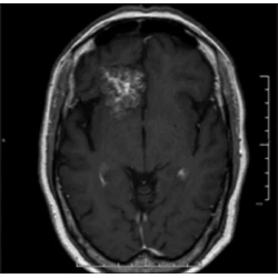
Andi Sadayandi Ramesh, Mahadevan Anita, Saini Jitender, Sampath Somanna DOI:10.4103/0976-3147.133582 PMID:25002766Medulloepithelioma is an extremely rare PNET in late adolescence and adults with only two cases noted in literature. These are WHO grade IV tumors with dismal prognosis. Only few cases survived beyond 5 months. We report a rare case of supratentorial medulloepithelioma in a 17 year old girl. She had presented with right sided weakness, headache and vomiting. Imaging showed an enhancing mass lesion in left parietal region which undergone gross total resection. After surgery, her headache, vomiting and right sided weakness improved. On histopathology, the tumor had characteristic trabecular, ribbon and palisaded arrangement with brisk mitotic activity, necrosis and calcification. Immuno-histochemistry revealed positivity for Synaptophysin, Vimentin and EMA while GFAP was negative. MIB-1 labeling was very high. Patient received postoperative radiotherapy. On follow up after 14 months, she was clinically asymptomatic with no recurrence on imaging. |
[ABSTRACT] [HTML Full text] [PDF] [Mobile Full text] [EPub] [PubMed] [Sword Plugin for Repository]Beta |
|
|
|
|
|
 |
Unilateral herpes zoster ophthalmicus with bilateral cerebral infarcts in human immunodeficiency virus seropositive patient |
p. 265 |
Girish Baburao Kulkarni, Veerendrakumar Mustare, Masoom Abbas Mirza DOI:10.4103/0976-3147.133584 PMID:25002767Ischemic stroke is a recognized complication of herpes zoster ophthalmicus. Arterial involvement is usually seen on the side of the rash. It is thought to be due to vessel inflammation by the virus, which travels from the trigeminal ganglion. Few case reports of bilateral and distant site of zoster lesions with stroke in the brain have been described. These reports suggest possibility of contiguous vascular, cerebrospinal fluid (CSF) or hematogenous spread of the virus from the ganglion to the cerebral blood vessels. Therapeutically acyclovir, anticoagulation, and steroids have been used in the treatment of the zoster associated with stroke. We describe a case of immunocompromised patient with ipsilateral zoster ophthalmicus with bilateral anterior circulation strokes, who was treated with above measures and made successful recovery. This report also raises/supports possible CSF/vascular/hematogenous spread of the virus from the ganglion to involve cerebral blood vessels leading to the stroke. |
[ABSTRACT] [HTML Full text] [PDF] [Mobile Full text] [EPub] [PubMed] [Sword Plugin for Repository]Beta |
|
|
|
|
|
 |
Spontaneous hemothorax associated with neurofibromatosis type I: A review of the literature |
p. 269 |
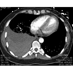
Swaroopa Pulivarthi, Byron Simmons, John Shearen, Murali Krishna Gurram DOI:10.4103/0976-3147.133592 PMID:25002768Neurofibromatosis is generally a benign disease, but has the potential for rare and fatal complications, such as spontaneous hemothorax. We report a case of massive hemothorax due to neurofibroma in a 49-year-old woman with neurofibromatosis type 1. The configuration of the radiological opacity and frank blood withdrawn on thoracentesis should suggest the diagnosis of hemothorax in a patient with neurofibromatosis. Surgical treatment for hemothorax is limited by arterial fragility and the prognosis is relatively poor. Any evidence of aneurysmal disease in the thoracic vessels should be aggressively managed percutaneously by coil embolization to prevent future rupture. |
[ABSTRACT] [HTML Full text] [PDF] [Mobile Full text] [EPub] [PubMed] [Sword Plugin for Repository]Beta |
|
|
|
|
|
|
COMMENTARY |
 |
|
|
|
Commentary |
p. 272 |
Keerati Hongsakul |
[HTML Full text] [PDF] [Mobile Full text] [EPub] [Sword Plugin for Repository]Beta |
|
|
|
|
|
|
CASE REPORTS |
 |
|
|
 |
All-trans-retinoic acid-induced pseudotumor cerebri in acute promyelocytic leukemia |
p. 273 |
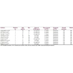
T. M. Anoop, Nidhi Jain, Sreejith G. Nair, Geetha Narayanan DOI:10.4103/0976-3147.133595 PMID:25002770All-trans-retinoic acid is an integral part in the treatment strategy of acute promyelocytic leukemia (APL). Here we describe a case of pseudotumor cerebri associated with all-trans-retinoic acid (ATRA) during the induction therapy in an adult with acute promyelocytic leukemia (APL). |
[ABSTRACT] [HTML Full text] [PDF] [Mobile Full text] [EPub] [Citations (2) ] [PubMed] [Sword Plugin for Repository]Beta |
|
|
|
|
|
 |
Organophosphorus-induced extrapyramidal intermediate syndrome in an adolescent suicide attempt survivor |
p. 276 |
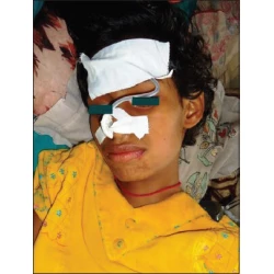
Sumantra Sarkar, Madhumita Nandi, Rakesh Mondal, Sandip Kumar Mandal DOI:10.4103/0976-3147.133596 PMID:25002771The victims of organophosphorus (OP) pesticide poisoning usually present with acute cholinergic crisis, due to the inhibition of the enzyme acetylcholinesterase. Any neurological complication in the form of intermediate syndrome is rare and its presentation with extrapyramidal symptoms is even rarer. The authors report such a case in a 12-year-old adolescent girl, who survived a near lethal suicidal attempt. |
[ABSTRACT] [HTML Full text] [PDF] [Mobile Full text] [EPub] [Citations (1) ] [PubMed] [Sword Plugin for Repository]Beta |
|
|
|
|
|
|
COMMENTARY |
 |
|
|
|
Commentary |
p. 278 |
Mostsfa Amr |
[HTML Full text] [PDF] [Mobile Full text] [EPub] [Sword Plugin for Repository]Beta |
|
|
|
|
|
|
Commentary |
p. 279 |
Ariful Basher |
[HTML Full text] [PDF] [Mobile Full text] [EPub] [Sword Plugin for Repository]Beta |
|
|
|
|
|
|
CASE REPORTS |
 |
|
|
 |
Olfactory region schwannoma: Excision with preservation of olfaction |
p. 281 |
Pravin Salunke, Devi Prasad Patra, Sameer Futane, Ritambhara Nada DOI:10.4103/0976-3147.133600 PMID:25002774Olfactory region schwannomas are rare, but when they occur, they commonly arise from the meningeal branches of the trigeminal nerve and may present without involvement of the olfaction. A 24 year old lady presented with hemifacial paraesthesias. Radiology revealed a large olfactory region enhancing lesion. She was operated through a transbasal with olfactory preserving approach. This manuscript highlights the importance of olfactory preservation in such lesions. |
[ABSTRACT] [HTML Full text] [PDF] [Mobile Full text] [EPub] [Citations (1) ] [PubMed] [Sword Plugin for Repository]Beta |
|
|
|
|
|
 |
A patient with pycnodysostosis presenting with seizures and porencephalic cysts |
p. 284 |
Subhash Kumar DOI:10.4103/0976-3147.133606 PMID:25002775Pycnodysostosis is a rare autosomal recessive disorder caused by mutations in the cysteine protease Cathepsin K gene located on chromosome 1q21. It has a well characterized skeletal phenotype which include short stature, generalized increased bone density with propensity of fractures, open calvarial sutures and fontanelle, dental abnormalities, obtuse mandibular angle, resorption of lateral end of clavicle, acro-osteolysis, and in some cases visceromegaly. Central nervous system involvement is very rare and porencephalic cysts has been reported only once, the cause being hypothesised to be an imbalance between the growing brain, its vascular supply and intraventricular fluid pressure. We had a patient with bilateral frontal lobe porencephalic cysts; the patient presenting with complex partial seizures. Cathepsins have been found to be involved in neurological diseases and role of proteases has been well established in gliosis. |
[ABSTRACT] [HTML Full text] [PDF] [Mobile Full text] [EPub] [Citations (3) ] [PubMed] [Sword Plugin for Repository]Beta |
|
|
|
|
|
 |
Hematogenous pasteurella haemolytica brain abscess |
p. 287 |
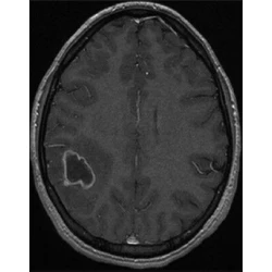
Mehdi Darmoul, Atef Ben Nsir, Mohamed Kilani, Mohamed Nejib Hattab DOI:10.4103/0976-3147.133608 PMID:25002776Pasteurella infections are common in domestic animals and very rare in human. We report a hematogenously acquired Pasteurella haemolytica brain abscess, mimicking brain tumor on magnetic resonance imaging, in an 18-year-old female patient known with cardiac interventricular communication, without recent history of animal contact. The outcome was good after abscess complete removal and antimicrobials therapy for 6 weeks. To the best of our knowledge, this is the first reported case of P. haemolytica brain abscess. |
[ABSTRACT] [HTML Full text] [PDF] [Mobile Full text] [EPub] [PubMed] [Sword Plugin for Repository]Beta |
|
|
|
|
|
 |
Hemichorea and dystonia due to frontal lobe meningioma |
p. 290 |
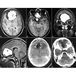
Abdul Qayyum Rana, Muhammad Saad Yousuf, Muhammad Zainuddin Hashmi, Zakerabibi Mohammed Kachhvi DOI:10.4103/0976-3147.133611 PMID:25002777Tumors originating from the meninges, also known as meningiomas, have rarely been known to cause parkinsonian symptoms and other movement disorders. Although some cases of AV malformations causing movement disorders have been described in the literature, not much has been reported about meningiomas in this regard. The aim of this case report is to further highlight the importance of brain imaging in patients with movement disorders for even a benign tumor; and also emphasize the need for a careful movement disorder examination because more than one phenomenology of movement disorders may result from the mechanical pressure caused by a tumor. We present a case report of a patient with a heavily calcified right frontal lobe meningioma. Our patient had irregular, involuntary, brief, fleeting and unpredictable movements of her left upper and lower extremities, consistent with chorea. The patient also had abnormal dystonic posturing of her left arm while walking. This case report highlights the importance of brain imaging as well as careful neurological examinations of patients with benign meningiomas. Moreover, it illustrates the remarkable specificity yet clinical diversity of meningiomas in presentation through movement disorders. |
[ABSTRACT] [HTML Full text] [PDF] [Mobile Full text] [EPub] [Citations (1) ] [PubMed] [Sword Plugin for Repository]Beta |
|
|
|
|
|
|
COMMENTARY |
 |
|
|
|
Commentary |
p. 292 |
George P Paraskevas |
[HTML Full text] [PDF] [Mobile Full text] [EPub] [Sword Plugin for Repository]Beta |
|
|
|
|
|
|
Commentary |
p. 293 |
Camila Catherine Aquino |
[HTML Full text] [PDF] [Mobile Full text] [EPub] [Sword Plugin for Repository]Beta |
|
|
|
|
|
 |
Cerebellar giant cell glioblastoma multiforme in an adult  |
p. 295 |
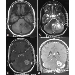
Sudhansu Sekhar Mishra, Sanjay Kumar Behera, Manmath Kumar Dhir, Satya Bhusan Senapati DOI:10.4103/0976-3147.133615 PMID:25002780Cerebellar glioblastoma multiforme (GBM) is a rare tumor that accounts for only 1% of all cases of GBM and its giant cell variant is even much rarely encountered in adults. A case of cerebellar giant cell GBM managed at our institution reporting its clinical presentation, radiological and histological findings, and treatment instituted is described. In conjunction, a literature review, including particular issues, clinical data, advances in imaging studies, pathological characteristics, treatment options, and the behavior of such malignant tumor is presented. It is very important for the neurosurgeon to make the differential diagnosis between the cerebellar GBM, and other diseases such as metastasis, anaplastic astrocytomas, and cerebellar infarct because their treatment modalities, prognosis, and outcome are different. |
[ABSTRACT] [HTML Full text] [PDF] [Mobile Full text] [EPub] [Citations (1) ] [PubMed] [Sword Plugin for Repository]Beta |
|
|
|
|
|
|
SHORT COMMUNICATIONS |
 |
|
|
|
Organophosphate intermediate syndrome with neurological complications of extrapyramidal symptoms in clinical practice  |
p. 298 |
Mark B. Detweiler DOI:10.4103/0976-3147.133616 PMID:25002781Organophosphates (OPs) are ubiquitous in the world as domestic and industrial agricultural insecticides. Intentional poisoning as suicides attempts are clinical phenomena seen in emergency departments and clinics in agricultural areas. Intermediate syndrome with the neurological complication of extra pyramidal symptoms following acute OP ingestion may occur in pediatric and adult cases. While death is the most serious consequence of toxic OP doses, low levels of exposure and nonfatal doses may disrupt the neurobehavioral development of fetuses and children in addition to bring linked to testicular cancer and male and female infertility. These are disturbing. Chronic and acute toxicity from OPs are barriers to the health of our present and future generations. Symptoms and treatment of acute and chronic OP exposure are briefly referenced with inclusion of the intermediate syndrome. Suggestions for local and systemic reduction of the acute and long term consequences of OP ingestion are opined. |
[ABSTRACT] [HTML Full text] [PDF] [Mobile Full text] [EPub] [Citations (1) ] [PubMed] [Sword Plugin for Repository]Beta |
|
|
|
|
|
|
IMAGES IN NEUROSCIENCES |
 |
|
|
 |
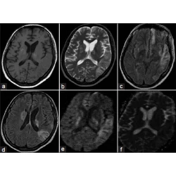
Frontocallosal leucoencephalopathy in subacute sclerosing panencephalits |
p. 302 |
CJ Suresh Chandran DOI:10.4103/0976-3147.133618 PMID:25002782 |
[HTML Full text] [PDF] [Mobile Full text] [EPub] [PubMed] [Sword Plugin for Repository]Beta |
|
|
|
|
|
|
LETTERS TO THE EDITOR |
 |
|
|
 |
Dreaded complications of mistaken identity - Hygroma vs effusion following decompressive craniotomy |
p. 305 |
Omekareswar Rambarki, Alugolu Rajesh DOI:10.4103/0976-3147.133623 PMID:25002783 |
[HTML Full text] [PDF] [Mobile Full text] [EPub] [PubMed] [Sword Plugin for Repository]Beta |
|
|
|
|
|
|
COMMENTARY |
 |
|
|
|
Commentary |
p. 307 |
Bhawna Sharma, Raghavendra Bakki Sannegowda, Parul Dubey |
[HTML Full text] [PDF] [Mobile Full text] [EPub] [Sword Plugin for Repository]Beta |
|
|
|
|
|
|
Commentary |
p. 308 |
Christian von der Brelie, Johannes Lemcke |
[HTML Full text] [PDF] [Mobile Full text] [EPub] [Sword Plugin for Repository]Beta |
|
|
|
|
|
|
Commentary
|
p. 309 |
Guru Dutta Satyarthee |
[HTML Full text] [PDF] [Mobile Full text] [EPub] [Sword Plugin for Repository]Beta |
|
|
|
|
|
|
LETTER TO THE EDITOR |
 |
|
|
 |
Adult onset subacute sclerosing panencephalitis - Lessons learnt from an atypical presentation |
p. 310 |
Mahendra Javali, Ramshekar Menon, Rahul Chakor DOI:10.4103/0976-3147.133637 PMID:25002787 |
[HTML Full text] [PDF] [Mobile Full text] [EPub] [Citations (1) ] [PubMed] [Sword Plugin for Repository]Beta |
|
|
|
|
|
|
COMMENTARY |
 |
|
|
|
Commentary |
p. 313 |
Adrią Arboix |
[HTML Full text] [PDF] [Mobile Full text] [EPub] [Sword Plugin for Repository]Beta |
|
|
|
|
|
|
LETTER TO THE EDITOR |
 |
|
|
|
Catatonia in a patient with bipolar disorder type I |
p. 314 |
Ather Muneer DOI:10.4103/0976-3147.133652 PMID:25002789 |
[HTML Full text] [PDF] [Mobile Full text] [EPub] [Citations (1) ] [PubMed] [Sword Plugin for Repository]Beta |
|
|
|
|
|
|
COMMENTARY |
 |
|
|
|
Commentary |
p. 317 |
Jurjen J. van Zwieten, Jan N. M. Schieveld |
[HTML Full text] [PDF] [Mobile Full text] [EPub] [Sword Plugin for Repository]Beta |
|
|
|
|
|
|
Commentary
|
p. 318 |
Walter Jaimes-Albornoz |
[HTML Full text] [PDF] [Mobile Full text] [EPub] [Sword Plugin for Repository]Beta |
|
|
|
|
|
|
Commentary |
p. 319 |
Roshan Bhad |
[HTML Full text] [PDF] [Mobile Full text] [EPub] [Sword Plugin for Repository]Beta |
|
|
|
|
|
|
LETTER TO THE EDITOR |
 |
|
|
|
Life threatening intracranial hemorrhages in malaria |
p. 320 |
Khichar Shubhakaran DOI:10.4103/0976-3147.133657 PMID:25002793 |
[HTML Full text] [PDF] [Mobile Full text] [EPub] [PubMed] [Sword Plugin for Repository]Beta |
|
|
|
|
|
|
COMMENTARY |
 |
|
|
|
Commentary |
p. 321 |
Jose Eymard Homem Pittella |
[HTML Full text] [PDF] [Mobile Full text] [EPub] [Sword Plugin for Repository]Beta |
|
|
|
|
|
|
LETTER TO THE EDITOR |
 |
|
|
|
Spasmodic dysphonia like presentation of stiff person syndrome |
p. 322 |
Abdul Qayyum Rana, Mohamed Sufian Masroor, Beenish Ismail DOI:10.4103/0976-3147.133659 PMID:25002795 |
[HTML Full text] [PDF] [Mobile Full text] [EPub] [PubMed] [Sword Plugin for Repository]Beta |
|
|
|
|
|
|
Accessible knowledge for tomorrow's surgeons and doctors |
p. 323 |
Thomas I. Lemon, Drew Davies DOI:10.4103/0976-3147.133661 PMID:25002796 |
[HTML Full text] [PDF] [Mobile Full text] [EPub] [PubMed] [Sword Plugin for Repository]Beta |
|
|
|
|
|