Generic selectors
Exact matches only
Search in title
Search in content
Post Type Selectors
Search in posts
Search in pages
Filter by Categories
Book Review
Brief Report
Case Letter
Case Report
Case Series
Commentary
Current Issue
Editorial
Erratum
Guest Editorial
Images
Images in Neurology
Images in Neuroscience
Images in Neurosciences
Letter to Editor
Letter to the Editor
Letters to Editor
Letters to the Editor
Media and News
None
Notice of Retraction
Obituary
Original Article
Point of View
Position Paper
Review Article
Short Communication
Systematic Review
Systematic Review Article
Technical Note
Techniques in Neurosurgery
View/Download PDF

Translate this page into:

Images in Neurosciences
10 (
1
); 125-125
doi:
10.4103/jnrp.jnrp_252_18

Os Odontoideum

Department of Neurosurgery, Hospital of Sion, CHVR, Sion, Switzerland
Address for correspondence: Dr. Arthur Robert Kurzbuch, Hôpital du Valais – Centre Hospitalier du Valais Romand (CHVR), Hôpital de Sion, Service de Neurochirurgie, Avenue du Grand-Champsec 80, CH-1951 Sion, Switzerland. E-mail: kurzbuch@web.de
Licence

This is an open access journal, and articles are distributed under the terms of the Creative Commons Attribution-NonCommercial-ShareAlike 4.0 License, which allows others to remix, tweak, and build upon the work non-commercially, as long as appropriate credit is given and the new creations are licensed under the identical terms.

Disclaimer:
This article was originally published by Medknow Publications & Media Pvt Ltd and was migrated to Scientific Scholar after the change of Publisher.

A 73-year-old patient presented to the neurosurgery clinic with a 2-year history of progressive paresthesia and deterioration of the dexterity of his right hand.

Neurologic examination revealed an impairment of fine motor skills of both hands and spastic hypertonus of the lower extremities. Computed tomography of the cervical spine showed a smooth-margined, independent osseous segment separated from the hypoplastic odontoid peg consistent with os odontoideum [Figure 1a, arrow].

Figure 1
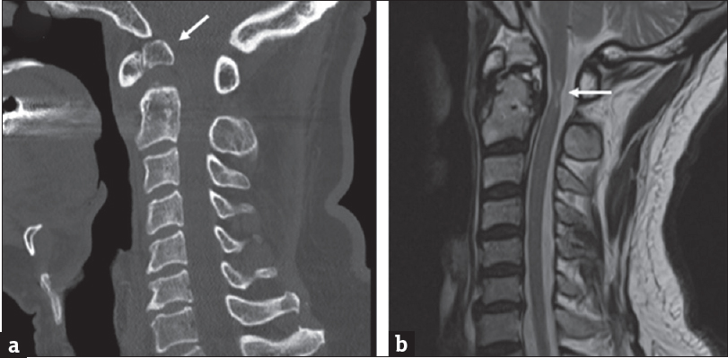
(a) Sagittal computed tomography of the cervical spine shows a separate odontoid process (arrow) from the body of C2. (b) Sagittal T2-weighted cervical magnetic resonance imaging reveals narrowing and hyperintensity (arrow) of the spinal cord at C2 level

The etiology of os odontoideum remains controversial with traumatic or congenital hypothesis.[12] It is incidentally discovered or seen in symptomatic patients with cervical myelopathy because of C1–C2 instability and spinal cord compression.[12] In this patient, magnetic resonance imaging revealed narrowing and hyperintensity of the spinal cord at C2 level on the T2-weighted sequence [Figure 1b, arrow]. C1–C2 posterior screw-rod stabilization was performed. Within 2 months after surgery, paresthesia had resolved, and the patient was able to write again.

Declaration of patient consent

The authors certify that they have obtained all appropriate patient consent forms. In the form, the patient has given his consent for his images and other clinical information to be reported in the journal. The patient understands that name and initials will not be published and due efforts will be made to conceal identity, but anonymity cannot be guaranteed.

Financial support and sponsorship

Nil.

Conflicts of interest

There are no conflicts of interest.

REFERENCES

  1. , , , . Os odontoideum: Etiology and surgical management. Neurosurgery. 2010;66:22-31.
    [Google Scholar]
  2. , , , . Incidental os odontoideum: Current management strategies. Neurosurg Focus. 2011;31:E10.
    [Google Scholar]
Show Sections