
Translate this page into:
Dermoid Cysts of the Anterior Fontanel in Adults
This is an open access journal, and articles are distributed under the terms of the Creative Commons Attribution-NonCommercial-ShareAlike 4.0 License, which allows others to remix, tweak, and build upon the work non-commercially, as long as appropriate credit is given and the new creations are licensed under the identical terms.
This article was originally published by Medknow Publications & Media Pvt Ltd and was migrated to Scientific Scholar after the change of Publisher.
Sir,
Dermoid cysts of anterior fontanel in adults are rare. These are benign cystic lesions that occur at lines of fusion in the body. Dermoid cysts of the scalp including at the anterior fontanel in infants and small children have been reported in the literature.[123] But not many reports exist on this condition in adults. Here, we present three adults who presented with this condition. The first patient was a 27-year-old young man who noticed this swelling about 7 years before. He noticed this when he started getting hurt while combing his hair. Clinical examination [Figure 1] revealed a well-rounded swelling of about 3 cm × 3 cm size in the region of the anterior fontanel. There were no visible pulsations, any sinus tract or punctum or dilated veins seen. On examination, the lesion had heterogeneous consistency with some areas soft and cystic. It was nontender and nonpulsatile and did not give a cough impulse. Transillumination was not present and there was no bruit on auscultation. The bony margins were felt to be inverted. The next patient was an 18-year-old boy who underwent tonsuring of the head and noticed the swelling of around 1.5 cm with a punctum. The other findings were similar. There was no discharge from the punctum. In this 22-year-old young female, the beautician noticed this swelling with similar findings but no punctum. All the patients underwent computed tomography (CT) scans which showed no intracranial extension in any case. Magnetic resonance imaging (MRI) was done in all cases and intracranial extensions were ruled out. All the patients underwent total excision of the lesion. During surgery, the lesions had hair and calcified portions and one of the swellings showed rudimentary teeth. There was some erosion of the outer table in one case (the young man), but there was no breach of the inner table in any of the cases. The histopathological report in all cases was dermoid cyst. Dermoid cysts in the head and neck are usually considered congenital lesions. However, occasionally, these lesions have been identified in adults also. The importance of dermoids in the scalp is the potential for intracranial extension. Here, we describe three adults with histologically proved dermoid cysts in the anterior fontanel region without any intracranial extension.
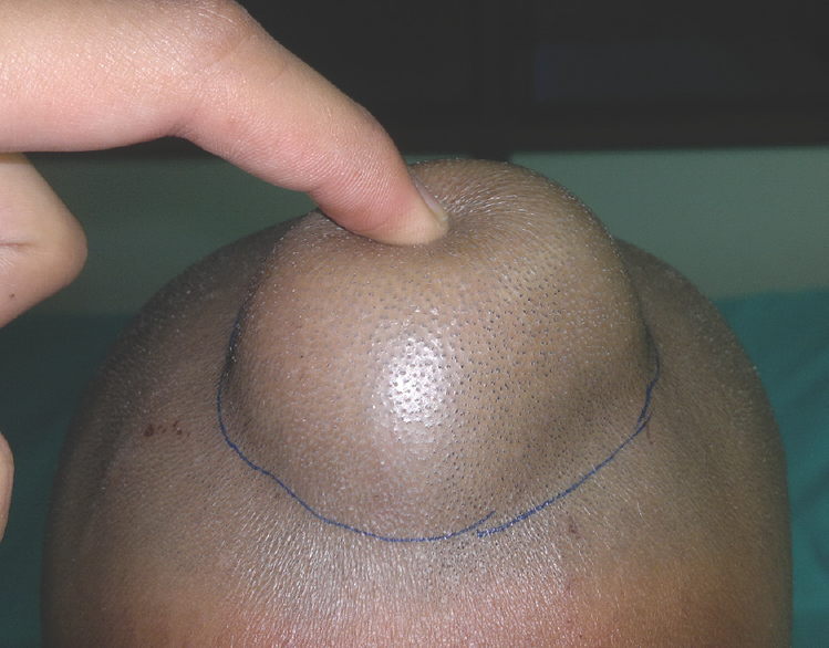
- Photograph of one male patient showing the dermoid cyst
de Carvalho et al.[3] presented 7 patients aged 3 months to 16 years with cystic lesions of the anterior fontanel. They were detected at birth and were progressive in size but asymptomatic. All patients underwent removal of the lesion. Ojikutu and Mordi[2] described congenital inclusion dermoid cysts over the region of the anterior fontanel in two adult Nigerians. Both were seen at birth but were not treated till adult life. There was no intracranial extension in both. Histopathologically, both were confirmed as dermoid cysts. Surgical excision was curative. de Castro et al.[4] also presented a 23-year-old man with a scalp swelling at the region of the anterior fontanel without any neurological issues. CT scan did not show any intracranial extension. The lesion was excised in total with very good result. The lesion was soft cystic containing a greenish viscous liquid with hair. Chaudhari et al.[5] described 21 children with congenital inclusion cysts of the subgaleal space. A majority of them were in the anterior fontanel. The cysts were reported as dermoid/epidermoid cysts. There have been many other studies on dermoids[67] over the anterior fontanel with similar conclusions. Reissis et al.[8] studied the histology of 16 craniofacial cysts and concluded that histological properties of dermoid cysts are conserved between craniofacial sites (periorbital, nasal, scalp, and postauricular). This reflects the consistency of ectodermal inclusion during early embryological development, which is independent of specific craniofacial site or surrounding anatomical structures.
In our study also, all the patients had neurologically asymptomatic lesions without any intracranial extension. All the lesions were confirmed to be dermoids histopathologically. All the lesions were excised totally without problems. The importance of these lesions for the surgeon is that these can mimic other pathologies such as encephalocoels or meningocoels, and so, CT scan/MRI is a must to rule out the pathology as well as any intracranial extension before any surgical intervention. The existence of this entity is especially useful for the trainee surgeons and neurosurgeons.
Declaration of patient consent
The authors certify that they have obtained all appropriate patient consent forms. In the form the patient(s) has/have given his/her/their consent for his/her/their images and other clinical information to be reported in the journal. The patients understand that their names and initials will not be published and due efforts will be made to conceal their identity, but anonymity cannot be guaranteed.
Financial support and sponsorship
Nil.
Conflicts of interest
There are no conflicts of interest.
REFERENCES
- Midline dermoid cyst resulting in frontal bone erosion. J Craniofac Surg. 2012;23:131-4.
- [Google Scholar]
- Congenital inclusion dermoid cyst located over the region of the anterior fontanel in adult Nigerians: Report of two cases. J Neurosurg. 1980;52:724-7.
- [Google Scholar]
- Dermoid cysts of the anterior fontanelle in adults: Case report. Arq Neuropsiquiatr. 2007;65:170-2.
- [Google Scholar]
- Congenital inclusion dermoid cysts of the scalp. Australas J Dermatol. 1992;33:135-40.
- [Google Scholar]
- Rapidly enlarging dermoid cyst over the anterior fontanel: A case report and review of the literature. Childs Nerv Syst. 2000;16:875-8.
- [Google Scholar]
- Craniofacial dermoid cysts: Histological analysis and inter-site comparison. Yale J Biol Med. 2014;87:349-57.
- [Google Scholar]


