
Translate this page into:
Kissing naevus arising from neural crest cells presenting as upper and the lower lid mass
This is an open-access article distributed under the terms of the Creative Commons Attribution-Noncommercial-Share Alike 3.0 Unported, which permits unrestricted use, distribution, and reproduction in any medium, provided the original work is properly cited.
This article was originally published by Medknow Publications & Media Pvt Ltd and was migrated to Scientific Scholar after the change of Publisher.
Abstract
A kissing nevus is a type of congenital compound nevus that affects equal portions of the upper and lower eyelid, and it extends to the lid margins. Congenital divided nevi of the eyelids are a rare melanocytic lesion. Only 30 patients are reported in the literature. We report a 40-year-old female of rural background who presented with a large painless enlarging pigmented mass, involving both upper and lower left eyelid since the past 20 years. Complete excision of the lesion was done, and the mass sent for histopathology, which revealed a compound nevus involving both lids. Surgery removed the obstruction, which had caused decreased visual acuity and had altered the cosmetic appearance of the patient. Lesions on the upper lid cause a mechanical ptosis, covers the visual axis, which causes obstruction in vision and is cosmetically unacceptable. There is a definite risk of malignant change in the nevus giving rise to malignant melanoma, and hence they should be removed as early as possible to give better functional and cosmetic results.
Keywords
Eyelid
melanoma
nevi

Introduction
A kissing nevus is a type of congenital compound nevus that affects equal portions of the upper and lower eyelid. Owing to its extension to the lid margins, the edges of the tumour touch or “kiss” during closure of the lids.[1] The kissing nevus origin dates to melanocyte migration or Schwann cells of a neuroectodermic origin during the embryological fusion of the lids at the 9th week of gestation, producing the “kissing” or split nevus.[2] The divided nevus is thought to arise during fetal development at a time when the eyelids are fused.[3] The lids appear as ectodermal protrusions from the 6th week of fetal development. They grow towards each other and gradually fuse until about the 24th week when they gradually separate. Melanoblasts originate from the neural crest and migrate during the 12–14 weeks stage of gestation to colonize the epidermis.
Case Report
A 40-year-old female presented to the eye OPD with a painless enlarging swelling both upper and lower left eyelid since the past 20 years [Figure 1]. History of trauma to the left eye with a stone was present 5 years back. The patient belonged to a rural background and low socioeconomic status. The patient never seeked medical intervention. The upper lid lesion was a soft tissue mass 2.5 cm × 0.5 cm × 0.5 cm. The skin over the lesion was pigmented with fine hair present over the mass. The lower eyelid mass was 2 cm × 0.8 cm × 0.5 cm, pigmented and hair bearing. Visual acuity (VA) in the right eye was 6/36 and left eye was hand movements close to face. Fundus of the right eye was normal and left eye media haze grade 4 was present. Slit lamp examination revealed adherent leucoma and early cataractous changes in the left eye. Cortical cataract was present in the right eye. Computed tomography scan revealed a heterogenous mass in the upper and lower lid in the left eye with extension to the median canthus. A surgery was planned where canthotomy, cantholysis were done, and lid repair in both the lids was carried out by the direct suturing method. The mattress suture was first passed at the grey line, and two more mattress sutures were padded one anterior and one posterior to the grey line. The inferior incision line was marked at 5 mm from the lash, and the upper line was drawn at the margin of the nevus. The entire upper eyelid, including the nevus, was excised, and a 5-mm-wide strip of orbicularis muscle and fat were removed. The tarsal plate and the skin of the eyelid were sutured using 6–0 vicryl to form a double-eyelid line. The skin muscle lamina was sutured with black silk. To remove the nevus in the lower eyelid, a full-thickness excision was performed. The lateral area of the anterior part of the eyelid of the orbicularis oculi muscle was anchored to the periosteum at the lateral orbital tubercle with interrupted sutures using 6–0 vicryl to provide additional support to the lower eyelid.
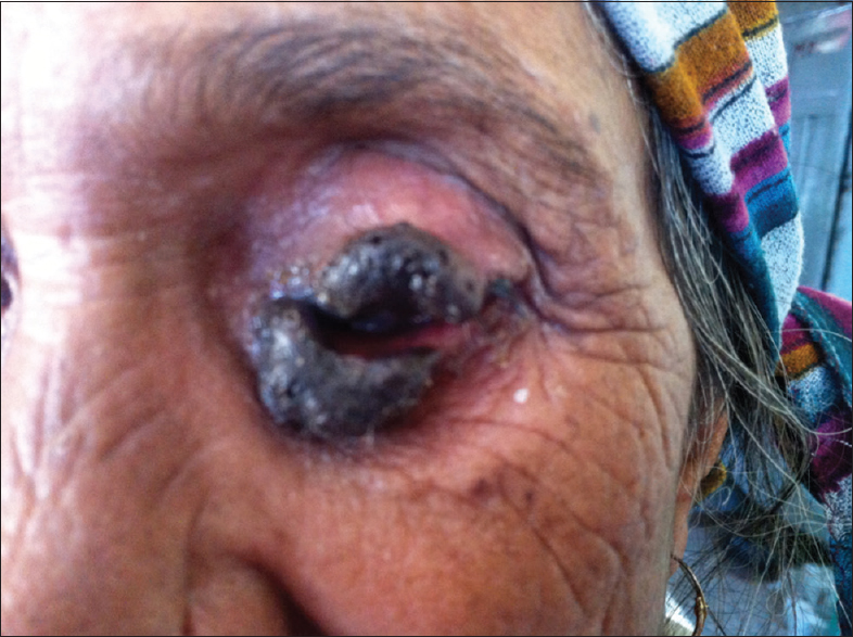
- Kissing nevus involving upper and lower lids
Histopathological examination of the section showed stratified squamous epithelium showing nests of nevus cells in dermal – epidermal junction. Upper dermis showed nests of round to cuboidal cells with vesicular nuclei, conspicuous nucleoli, moderate cytoplasm with abundant melanin in the stroma. Mid dermis had cords of nevus cells with scant cytoplasm. Histopathological report suggested of a melanocytic compound nevus [Figure 2]. The patient was kept under observation for 4 days, antiseptic dressing was done [Figure 3]. No postoperative complication was present. On her follow-up visit on 10.10.2014 her skin over the lids was normal and healed and VA improved to 6/36 in the left eye [Figure 4].

- Histopathology of the lesion revealing a compound naevus

- Photograph of the patient on 1st postoperative day

- Postoperative photograph after 2 months
Discussion
Congenital nevi occur in approximately 1% of all newborns, with the vast majority being <1.5 cm in size.[4] Nevocellular nevi are benign proliferations of melanocytic cells divided into 3 categories depending on their architecture: Junctional, compound and intradermal compound nevi possess features of junctional (arising from the deeper layers of the epidermis or “junctional region”) and intradermal nevi.
On the eyelid, the nevus often arises at the eyelid margin and may be flat, elevated, dome-shaped, and even pedunculated. The flat lesions are often junctional nevi. The domeshaped lesions are often intradermal, or compound nevi, and the pedunculated lesions are usually intradermal nevi. Generally nevi will be tan in color and often will feature deep brown pigmentation. The nevus is generally well circumscribed and not associated with ulceration.
Congenital divided nevus of the eyelids was first described in 1919 by Fuchs.[5] Since then about 30 cases have been reported. In 1937, Collenza reported on two patients with divided naevi: A 19-year-old girl and a 26-year-old man, Callahan, and Harrison and Okun reported single cases. The most recent series was reported by Ehlers in 1969. He described 10 cases, the youngest patient being a 15-year-old boy: Two patients had simple excision, two needed full-thickness skin grafts to repair the defect, two were treated with cryotherapy, and the rest received no treatment.
Suspicious pigmented lesions of the eyelid, which have a history of growth, alteration in pigment pattern, vascularity, associated inflammation should be removed as the early diagnosis of melanoma is critical and may be confused with a nevus. Some nevi in which the diagnosis is obvious may be cosmetically unacceptable to the patient or may cause irritation, particularly if they are pedunculated. The lifetime risk of malignant degeneration in small congenital nevi is not clearly established. Large cutaneous melanocytic nevi (>4 cm), however, do give rise to melanoma.[6] The risk of malignant transformation is 4.6% during a 30 years period.[7]
Dermabrasion is a successful technique for removing congenital melanocytic nevus if the lesion is treated early in life and the nevus cells are still limited to the superficial dermis.[8] If the nevus involves the deep dermis and subcutaneous tissue, then the treatment consists of full-thickness excision followed by repair with full-thickness skin graft or split skin.
Prognosis
There is a certain risk of malignant change in the nevus giving rise to malignant melanoma, which is more documented for large nevi, but is thought to exist for medium sized nevi as well.[9] The reported incidence of malignant change is very variable, ranging from 2% to 30% depending on the length of follow-up, with an average of 14% for a whole lifetime. A melanocytic nevus involving the eyelids may affect visual development if the mass on the upper lid causes a mechanical ptosis and covers the visual axis.[10]
Conclusion
Kissing nevi of the eyelids may be cosmetically unacceptable causing depression and a feeling of low confidence. It can cause functional problems including ptosis and visual field defects. Management requires surgical excision and reconstruction. Initial, complete excision is important because residual tumour can grow, often with a more verrucous or thickened appearance making subsequent determination of malignant transformation and reconstruction difficult for the ophthalmologists.
Source of Support: Nil.
Conflict of Interest: None declared.
References
- Histochemistry and development of the human eyelids. Acta Ophthalmol (Copenh). 1965;43:642-68.
- [Google Scholar]
- Periocular congenital melanocytic nevi. J Pediatr Ophthalmol Strabismus. 1986;23:222-6.
- [Google Scholar]
- The incidence of malignant transformation in giant pigmented nevi. Scand J Plast Reconstr Surg. 1977;11:163-7.
- [Google Scholar]
- Permanent removal of pigmentation from giant hairy naevi by dermabrasion in early life. Br J Plast Surg. 1977;30:321-3.
- [Google Scholar]
- The risk of malignancy in large congenital nevi. Plast Reconstr Surg. 1974;53:421-8.
- [Google Scholar]
- Giant hairy nevus: Preventable cause of amblyopia. J Pediatr Ophthalmol. 1976;13:192-5.
- [Google Scholar]


