
Translate this page into:
A case of Ross syndrome presented with Horner and chronic cough
This is an open-access article distributed under the terms of the Creative Commons Attribution-Noncommercial-Share Alike 3.0 Unported, which permits unrestricted use, distribution, and reproduction in any medium, provided the original work is properly cited.
This article was originally published by Medknow Publications & Media Pvt Ltd and was migrated to Scientific Scholar after the change of Publisher.
Abstract
Ross syndrome is a rare sweating disorder associated with Adie's tonic pupil, decreased or diminished tendon reflex and unknown etiology. Although autonomic disturbances affecting sudomotor and vasomotor functions are seen commonly, they are rarely symptomatic. While Ross syndrome is typically characterized with dilated tonic pupil, it may be rarely manifested with miotic pupils (little old Adie's pupil), which can make diagnosis difficult. In this article, we aim to specify the atypical clinical manifestations of syndrome by means of Ross syndrome manifested by autonomic symptoms, Horner syndrome, chronic cough together with bilateral little old Adie's pupil.
Keywords
Chronic cough
hemihyperhidrosis
Horner syndrome
little old adie's pupil

Introduction
Ross syndrome is a progressive, degenerative, and autonomic nervous system disorder.[1] The disease comprises classical triad of Adie's tonic pupil, decreased or diminished tendon reflexes, and sweating disorders especially anhidrosis.[2] However, basic objective sign is hyperhidrosis, which is often revealed by a segmental compensatory.[3] Although other autonomic disturbances occur in addition to this, they are quite rarely symptomatic.[1] Hyperhidrosis in autonomic disturbances is expressed to be compensatory over-response of endocrine glands remained from postganglionic injury.[4] Chronic cough occurs as a result of efferent or afferent involvement of vagus nerve.[5] Horner's syndrome occurs in consequence of damage to superior cervical sympathetic autonomic ganglion and it is characterized by miosis, upper eyelid ptosis, loss of ciliospinal reflex on the affected side, and less commonly loss of sweating, which may extend from the face and neck to third rib and third thoracic spine on the same side.[26]
Case Report
A 48-year-old female patient presented with the complaint of excessive sweating in the right side of the body for 10 years. She said that her complaint increased much more during exercise and in hot weathers. She had no prior traumatic event, pyretic disease, or stroke before these complaints. Physical examination demonstrated flushing on the right half of the face, hyperhidrosis on the right face and neck, wetness on the upper region of right chest, shoulder, armpit, and upper region of the back of the dress. Neurological examination showed slight ptosis of the left eyelid, pupillary miosis more markedly on the left side and pupil irregularity on the left side. Deep tendon reflexes were absent in both upper and lower extremities. The other neurological functions were intact. The patient experienced cough attacks during the examination and it was learnt that this complaint was present for 5 years. In addition, it was learnt, during inquiry, that the patient had photophobia, blurred vision, and reading difficulty complaints for 2 years. Detailed examination and tests were planned for the patient.
Laboratory testing glucose, HbA1c, erythrocyte sedimentation rate, serum electrophoresis, autoantibody screen (Antinuclear Antibody, Rheumatoid Factor, anti-SSA, anti-SSB), antithyroid antibodies, syphilis serology (fluorescent treponemal antibody), Schirmer test were normal. Cranial, cervical, thoracic magnetic resonance imaging, and thoracic computed tomography revealed no abnormalities. At the electrophysiological investigation performed on the patient, motor and sensory nerve transmission studies of the upper and lower extremities were normal and bilateral, median, and tibial somatosensory evoked potential (SEP) investigations were normal. Sympathetic skin response (SSR) was obtained to be normal from the right upper and lower extremities but it could not be obtained from the left upper and lower extremities. Bilateral tibial H-reflex could not be obtained.
Sweat test was performed on the patient. She was unclothed and the entire body surface, except periorbital of face and genitalia, was covered with a 2% alcoholic solution of iodine and with rice starch powder. The color of rice starch powder changed from white to black in the presence of sweat. Digital pictures of subjects were taken. According to the sweat test, it was found to be hypohidrosis on the left face, neck, shoulder, and upper pectoral and anterior regions until T3-4 dermatome levels [Figure 1].
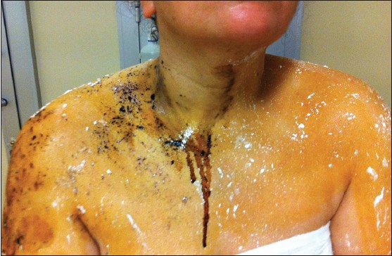
- Sweat test
During the ophthalmological examination of the patient, performed in the eye clinic, it was determined that pupils were bilaterally miotic with more markedly on the left side and irregular on the left side and bilateral direct and indirect light reflexes could not be obtained [Figure 2a]. Bilateral vision of the patient was complete and posterior segment examination was normal. During dilute pilocarpine test, miosis was not observed in both of the pupils. Slow and partial dilatation were observed in both the pupils during far looking and miosis was observed in both the pupils during near looking [Figure 2b and c]. On the other hand, dilatation was detected in the left eye and ptosis was partially resolved, with 1% phenylephrine test performed in the both the eyes.

- Patient's pupils (a) light reflex response, (b) pupils during far looking, (c) pupils during near looking
Additionally, noninvasive autonomic tests were performed on the patient. Orthostatic test may determine autonomic dysregulation.[7] It appears that the RR interval mode can also provide greater information in the heart rate variability responses to the change in blood pressure. Using current methods of analysis, it is also possible to gain a detailed look at the actual variability during the laying and then the standing periods. During orthostatic test of these tests, a reduction of 35 mmHg was determined in systolic blood pressure (normal value of reduction: <30 mmHg) and a reduction of 20 mmHg was determined in diastolic blood pressure (normal value of reduction: <15 mmHg). After a complete rest of 15 min in the supine position, the ECG recording was started and the subject assumed erect posture from the supine position as quickly as possible (within 3 s) with continuous electrocardiography recording for 30 s or more in erect posture. 30:15 ratio was calculated by taking the ratio of longest RR interval around 30th beat to shortest RR interval around 15th beat after standing. During 30:15 rate test (normal value of 30:15 rate: >1.07), 30:15 rate was determined to be 1.0. During Valsalva rate test (normal value of valsalva rate: >1.20), this rate was determined to be 1.0. So, we determined autonomic dysregulation in the patient.
It was learnt that the patient was examined in detail by chest diseases polyclinic 5-6 years ago due to chronic cough (thoracic computed tomography, pulmonary function tests, purified protein derivative (PPD) test, allergy panel, complete blood count) but no cause could be found. The cough was refractory to therapeutic trials of inhaled, nasal, and oral steroids, lignocaine spray and nebulizer, local anesthetic lozenges, and oral H2 antagonists. A trial of bornaprin HCl orally 4 mg/day was applied for compensatory hyperhidrosis. No adverse effect was observed except drug-related moderate dryness for 6 months and the symptom was partially decreased.
Discussion
Holmes-Adie syndrome is a neurological disorder characterized by a tonically dilated pupil (defined to be a dilated pupil exhibiting poor light response and light-near dissociation) and loss of deep tendon reflexes. Harlequin syndrome is characterized by loss of unilateral sudomotor activity without ocular sympathetic innervation. Combination of Harlequin's syndrome and Adie's syndrome is known to be Ross syndrome.[34] Ross syndrome, which was described for the first time in 1958, is a complex and progressive disorder of peripheral autonomic nervous system. Its exact pathogenesis is unknown.[89] There is a large engagement between Ross syndrome, Holmes–Adie syndrome and more common autonomic diseases both clinically and pathogenetically.[89] In some cases published, observation of some symptomatic and asymptomatic autonomic disturbances other than triad described (tonic pupil, loss of deep tendon reflexes, and segmental hypohidrosis) show that the disease is a dysautonomic disorder in which different reflexions occur due to widespread damage to ganglion cells and their extensions seen rather than to be a partial autonomic disease.[9] Hypohidrosis seen in these diseases are generally segmental and progressive and considered to be caused by damage to postganglionic sympathetic fibers innervating the sweat glands.[2]
Increase in cutaneous blood flow manifesting itself as flushing along with compensatory contralateral excessive sweating due to disorder in heat consumption in these patients increases in the conditions in which heat production increases like hot weather and excessive exercise.[38] In our case, hypohidrosis and compensatory contralateral excessive sweating was visible to inspection on face, neck, shoulders, and upper chest. We obtained results supporting these findings also in sweat test. Also, excessive sweating, which is the major disturbing complaint of the patient, increased much more with exercise and hot weather. In addition, not obtaining SSR response from the left upper and lower extremities supports compensatory hyperhidrosis hypothesis by establishing efferent dysfunction of somato-sympathetic reflex arc on the left side.
Pathophysiology of Adie's pupil is aberrant regeneration resulting from damage to parasympathetic cholinergic fibers between iris and ciliary ganglion.[2] Adie's pupil is classically described as tonic, dilated pupil with diminished or loss of light reflex. While Adie's pupil is dilated initially, it begins to constrict (miosis) progressively approximately after 2 years because of the aberrant reinnervation by accommodative fibers and this is called chronic tonic pupil or little old Adie's pupil. In this way, most of Adie's pupil can be confused with Argyl–Robertson's pupil, Horner syndrome and miotic pupil of elderly little pupil of elderly. In Ross syndrome, unique pupil is initially affected typically and shows tendency to be bilateral over the years. Therefore, anisocoria is quite typical both at the beginning and advancing times of the disease. In most of the patients with Ross syndrome, while some residual light reflex is seen in the affected pupil, iris sphincter is unresponsive in 10% of the patients. Parasympathetic innervation of the pupils was investigated through administration of a single drop of a dilute solution (0.125%) of pilocarpine in each eye. An absolute pupillary constriction of 1.5 mm or greater was considered suggestive of cholinergic supersensitivity.[6] Miosis may not be seen with diluted pilocarpine in the patients with little old Adie's pupil in some cases.[234] However, tonic and slow response of pupil to accommodative stimulus is most distinguishing feature for diagnosis. This condition is stated to be near-light dissociation.[24] Also, in our case, miosis was being observed in both of eyes (more markedly on the left side), light reflex was absent, slow tonic accommodative response to near-to-far was present and no response was observed to diluted pilocarpine test. Presence of Horner's syndrome was considered in the patient with ptosis of the left eyelid. Phenylephrine test was used for diagnosis and localizing the site of the lesion of Horner's syndrome.[1011] Dilatation response in left eye performed with 1% phenylephrine test showed that there was postganglionic adrenergic denervation supersensitivity (postganglionic Horner syndrome). Absence of light reflex showed that Adie's pupil and eye findings of Horner's syndrome were concomitantly present in the left eye. In the published cases, dilated pupils showing light-near dissociation were described as it was known classically but no case showing association with bilateral miotic pupil and Horner's syndrome was encountered like in our case.[611]
Hyporeflexia in Ross syndrome is progressive, bilateral, and symmetrical. Although its etiology is not clear, it is estimated to be caused by presynaptic degeneration of dorsal root fibers transmitting impulse to anterior horn cells.[2] Also, in our case, deep tendon reflexes could not be obtained commonly. This condition was shown electrophysiologically by not obtaining bilateral tibial H-reflexes.
Cough can be related to damage to afferent or efferent fibers of vagus nerve and it is also rarely encountered in diseases causing autonomic dysfunctions. There are a few reports of Ross syndrome associated with cough in the literature.[5]
Systemic anticholinergic drugs, topical medications, administration of botulinum toxin, treatment with modified iontophoretic device, and the surgical treatments like thoracic sympathectomy were tried for hyperhidrosis.[12131415] Treatment decision is very important because compensatory hyperhidrosis treatment may lead to heat exhaustion and heat stroke. Heat intolerance may be managed by wearing wet clothing during physical activity in order to prevent hyperthermia and hyperhidrosis to some extent.
Our case may show that the Ross syndrome is not a limited autonomic disturbance but a different clinical reflection of a common autonomic disturbance and the syndrome may be presented with different clinical manifestations particularly as pupil symptoms based on the disease age.
Source of Support: Nil.
Conflict of Interest: None declared.
References
- Cardiovascular and sweating dysfunction in patients with Holmes-Adie syndrome. J Neurol Neurosurg Psychiaty. 1993;56:1096-102.
- [Google Scholar]
- Bilateral tonic pupils: Holmes Adie syndrome or generalised neuropathy? Br J Ophthalmol. 2007;91:1620-3.
- [Google Scholar]
- Ross syndrome with sweating anomaly associated with Sjögren syndrome: An infrared thermo-graphic case study. Acta Derm Venereol. 2011;91:80-1.
- [Google Scholar]
- Neuro-ophthalmic diagnoses. In: Levin LA, Arnold AC, eds. Neurophtalmology the Practical Guide. New York: Thieme Medical Publishers; 2005. p. :325-40.
- [Google Scholar]
- Chronic cough in the Holmes-Adie syndrome: Association in five cases with autonomic dysfunction. J Neurol Neurosurg Psychiatry. 1998;65:583-6.
- [Google Scholar]
- Ocular drugs in clinical practice. In: Holdeman NR, ed. Clinical Ocular Pharmacology (5th ed). St Louis: Butterworth-Heinemann, Elsevier; 2008. p. :349-81.
- [Google Scholar]
- The relationship between orthostatic dysregulation and the orthostatic test in dizzy patients. Eur Arch Otorhinolaryngol. 1996;253:268-72.
- [Google Scholar]
- Ross syndrome: A rare or a misknown disorder of thermoregulation. A skin innervation study on 12 subjects? Brain. 2006;129:2119-31.
- [Google Scholar]
- Ross syndrome plus: Beyond Horner, Holmmes-Adie, and Harlequin. Neurology. 2000;55:1841-6.
- [Google Scholar]
- Segmental anhidrosis with hyporeflexia associated with congenital spinal deformity: A Ross's syndrome variant or inverse Horner's syndrome? Indian J Dermatol Venereol Leprol. 2004;70:29-32.
- [Google Scholar]
- Pharmacologic testing in Horner's syndrome-a new paradigm. S Afr Med J. 2010;100:738-40.
- [Google Scholar]
- Treating hyperhidrosis. Anticholinergic drugs were not mentioned. BMJ. 2000;321:703.
- [Google Scholar]
- Use of topical glycopyrrolate in Ross syndrome. J Am Acad Dermatol. 2006;55(Suppl 5):S111-2.
- [Google Scholar]
- Selective degeneration of sudomotor fibres in Ross syndrome and successful treatment of compensatory hyperhidrosis with botulinum toxin. Muscle Nerve. 1998;21:1790-3.
- [Google Scholar]
- Ross syndrome: Treatment of segmental compensatory hyperhidrosis by a modified iontophoretic device. J Am Acad Dermatol. 1993;28:308-12.
- [Google Scholar]


