
Translate this page into:
Crossed cerebellar atrophy
This is an open-access article distributed under the terms of the Creative Commons Attribution-Noncommercial-Share Alike 3.0 Unported, which permits unrestricted use, distribution, and reproduction in any medium, provided the original work is properly cited.
This article was originally published by Medknow Publications & Media Pvt Ltd and was migrated to Scientific Scholar after the change of Publisher.
A 20-year-old male presented with history of left focal seizures since 10 years. At the age of 10 years, he suffered from an encephalitic illness with focal seizures and was comatose for 2 days. Subsequently, he had residual left hemiparesis with left focal seizures. He used to get infrequent seizures once in 2–3 months on 600 mg of carbamazepine and 300 mg of phenytoin.
On examination, he was conscious and alert. He had left Upper motor neuron facial weakness and left hemianopia on confrontation examination. Motor system exanimation showed left hemiparesis; Medical research council grade 3/5 with dystonic posturing of the left hand. There were no cerebellar signs
Magnetic resonance imaging scan of the brain showed severe atrophy of the right temporoparietal and occipital areas with exvacuo dilatation of the right lateral ventricle [Figure 1]. A well-defined cystic lesion suggestive of porencephalic cyst was seen in the right high parietal lobe with adjacent dystrophic calcification [arrow, Figure 1]. Left cerebellar hemisphere showed diffuse atrophy with prominent folia [Figure 2] with atrophy of the left middle cerebellar peduncle [arrow, Figure 3]. These finding are suggestive of crossed cerebellar atrophy (CCA).
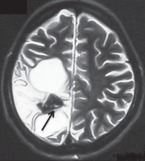
- Axial T2-weighted image showing atrophy of right temporoparietal and occipital areas with exvacuo dilatation of the right lateral ventricle. A porencephalic cyst in the right high parietal lobe with adjacent dystrophic calcification (arrow)

- Axial T2-weighted image showing left cerebellar atrophy with prominent folia

- Axial T2-weighted image showing left cerebellar atrophy with atrophy of the left middle cerebellar peduncle (arrow)
CCA is the atrophy of the cerebellum contralateral to the supratentorial hemispheric lesion. Long-standing unilateral cerebral hemisphere lesions due to prenatal brain injury, postnatal hypoxic brain damage or stroke in adults can result in CCA. Proposed mechanisms of CCA include uncontrolled seizures leading to transneuronal degeneration and post-ictal damage.[1]
A study done on 51 adult patients with epilepsy and precocious destructive lesions found that extent of supratentorial lesion and status epilepticus are deciding variables for the development of CCA.[2] Crossed cerebellar diaschisis (CCD), seen as contralateral cerebellar hypometabolism on Positron emission tomography (PET) scan, has been described as transient reversible functional impairment in an area remote from the site of a primary lesion.[3]
Reversible CCD and irreversible CCD are probably the extremes of the same biological process. FLAIR image showed right parietal atrophy with ex vacuo ventricular dilatation with left cerebellar atrophy. [Figure 4]

- FLAIR image showing right parietal atrophy with ex vacuo ventricular dilatation with left cerebellar atrophy
Our patient had contralateralmiddle cerebellar peduncle atrophy suggesting damage to the corticopontinecerebellar pathway leading to transneuronal degeneration. Our patient is on regular follow-up with infrequent, once in 2–3 months, seizures on 800 mg of carbamazepine and 300 mg of phenytoin.
Source of Support: Nil
Conflict of Interest: None declared
References
- Crossed cerebellar atrophy: An old problem revisited. ActaNeuropathol. 1982;57:197-202.
- [Google Scholar]
- Crossed cerebellar atrophy in patients with precocious destructive brain insults. Arch Neurol. 2002;59:843-7.
- [Google Scholar]
- “Crossed cerebellar diaschisis” in human supratentorial infarction. Ann Neurol. 1980;8:128.
- [Google Scholar]


