
Translate this page into:
Cirsoid aneurysm of scalp
This is an open-access article distributed under the terms of the Creative Commons Attribution-Noncommercial-Share Alike 3.0 Unported, which permits unrestricted use, distribution, and reproduction in any medium, provided the original work is properly cited.
This article was originally published by Medknow Publications & Media Pvt Ltd and was migrated to Scientific Scholar after the change of Publisher.
A 40-year-old lady presented with swelling on the right side of the scalp associated with continuous headache for 1 month. Her general physical examination was normal. Local examination revealed a pulsatile swelling on the right temporal-occipital region of the scalp. Few dilated veins were noted in this region. On auscultation, a bruit was detected over the lesion. Other systemic examination was normal, including thyroid, breast, and musculoskeletal system.
Patient underwent computed tomography of the brain along with angiography. Aneurysmal dilated mass of vessels were detected in the right temporal-occipital region of scalp in the subcutaneous plane [Figure 1], which enhanced markedly. Feeding vessel was from the occipital artery posteriorly and the superficial temporal artery [Figure 2]. Large draining veins were noted running anteriorly and posterior to the occiput [Figure 3]. A diagnosis of cirsoid aneurysm of scalp was made. Patient was advised to undergo excision of the aneurysm, but the patient refused surgery. She was discharged with an advice of regular follow-up.
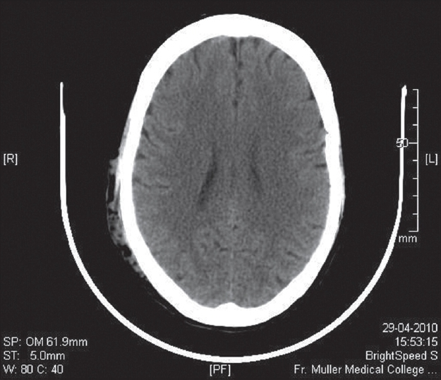
- Unenhanced axial CT image shows a serpiginous subcutaneous mass in the right temporal-occipital region
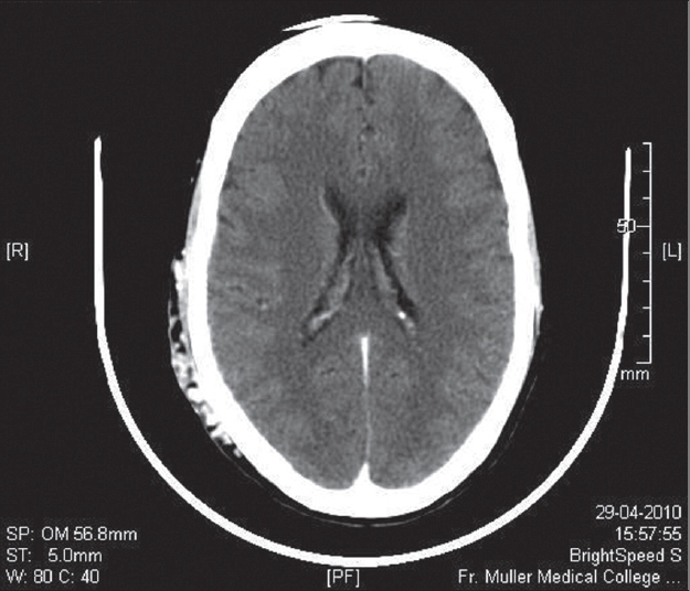
- Contrast-enhanced axial CT image shows marked contrast enhancement within the cirsoid aneurysm
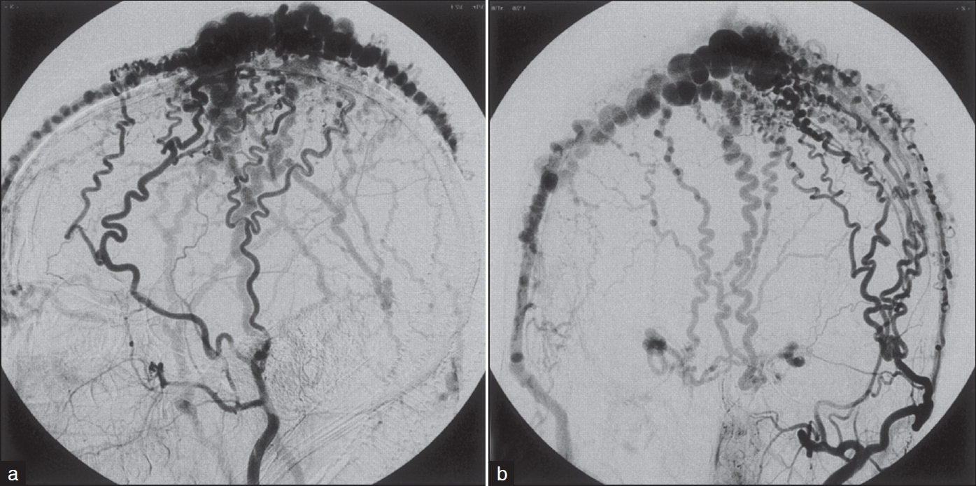
- CT angiogram showing the lesion
Cirsoid aneurysms of the scalp were first described in 1833 by Brecht.[1] Cirsoid aneurysms are rare arterio-venous fistulas of the scalp. They are usually congenital in etiology. However, traumatic fistulas have also been described.[1] In 90% of the patients, the superficial temporal artery is the main supply to the fistula with only one dominant feeding artery in 71% of patients.[2] In the remaining cases, there is usually involvement of both the superficial temporal and occipital arteries.[12] Untreated patients can develop progressive scalp and facial cosmetic deformity from the markedly tortuous subcutaneous vessels. However, this condition is not life-threatening.[2] Surgical resection of the fistula is usually successful. Endovascular and percutaneous occlusion of the fistulas have been described. However, the results have been mixed.
Differential diagnosis of a pulsatile scalp swelling includes the following: superficial temporal artery aneurysm although rare should be considered when a temporal region mass is evaluated. This condition is almost always a result of blunt or penetrating head trauma. Clinical examination with imaging confirms the diagnosis. Simple elective ligation and excision of the aneurysm are curative.[3]
A-V malformations may be congenital or traumatic in origin. Clinical manifestation may include a loud continuous bruit, hemorrhage and throbbing headache, and in severe cases scalp necrosis. Angiography is the investigation of choice. Primary lesions can be excised completely without significant blood loss and scalp necrosis.[4] Metastatic deposits from follicular carcinoma of thyroid have to be ruled out prior to the diagnosis of cirsoid aneurysm.
Source of Support: Nil.
Conflict of Interest: None declared.


