
Translate this page into:
Musculocutaneous nerve substituting for the distal part of radial nerve: A case report and its embryological basis
This is an open-access article distributed under the terms of the Creative Commons Attribution-Noncommercial-Share Alike 3.0 Unported, which permits unrestricted use, distribution, and reproduction in any medium, provided the original work is properly cited.
This article was originally published by Medknow Publications and was migrated to Scientific Scholar after the change of Publisher.
Abstract
In the present case, we have reported a unilateral variation of the radial and musculocutaneous nerves on the left side in a 64-year-old male cadaver. The radial nerve supplied all the heads of the triceps brachii muscle and gave cutaneous branches such as lower lateral cutaneous nerve of the arm and posterior cutaneous nerve of forearm. The radial nerve ended without continuing further. The musculocutaneous nerve supplied the brachioradialis, extensor carpi radialis longus and extensor carpi radialis brevis muscles. The musculocutaneous nerve divided terminally into two branches, superficial and deep. The deep branch of musculocutaneous nerve corresponded to usual deep branch of the radial nerve while the superficial branch of musculocutaneous nerve corresponded to usual superficial branch of the radial nerve. The dissection was continued to expose the entire brachial plexus from its origin and it was found to be normal. The structures on the right upper limb were found to be normal. Surgeons should keep such variations in mind while performing the surgeries of the upper limb.
Keywords
Anatomical variant
embryology
musculocutaneous nerve
radial nerve
upper limb

Introduction
In the upper limb, usually the musculocutaneous nerve branches out from the lateral cord of brachial plexus. It passes through the coracobrachialis muscle to innervate it and the biceps brachii muscle. Then, it supplies brachialis muscle and continues as the lateral cutaneous nerve of forearm without exhibiting any communication with the radial nerve or any other nerve.[1] The radial nerve is usually a continuation of the posterior cord of the brachial plexus. The radial nerve supplies all heads of the triceps brachii muscle and gives branches such as the lower lateral cutaneous nerve of the arm as well as the posterior cutaneous nerve of the forearm. Then, the radial nerve enters the anterior compartment of arm under cover of the brachioradialis muscle. Here, it supplies brachioradialis, extensor carpi radialis longus and extensor carpi radialis brevis muscles before it divides into superficial and deep branches. Its superficial branch becomes subcutaneous while the deep branch pierces supinator muscle and supplies the remaining muscles of extensor compartment of forearm.[1]
The brachial plexus variations and other variant innervation of the upper limb have been reported continuously since the 19th century.[2] The anatomical variations of the peripheral nerves are important to orthopedic surgeons, neurophysicians, physiotherapists and radiologists. Such comprehension is useful in nerve grafting and neurophysiological evaluation for diagnosing peripheral neuropathies.[3] In the present case report, we present unique unilateral variations of radial and musculocutaneous nerves.
Case Report
The present variation was observed during routine dissection at the anatomy department of a medical college of a rural hospital in central India. In a 64-year-old male cadaver, the unilateral variation in motor innervations of the left arm was observed. The radial and musculocutaneous nerves were found to as variants. The radial nerve supplied all the heads of the triceps brachii muscle and ended without continuing further. The lower lateral cutaneous nerve of arm was emerging from the lateral head of triceps brachii while the posterior cutaneous nerve of the forearm was emerging from the medial head of triceps brachii muscle [Figure 1].
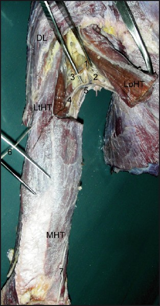
- Dissection showing the variation of radial nerve (DL: deltoid; LoHT, LtHT and MHT: Long, lateral and medial head of triceps brachii, respectively; 1: radial nerve; 2, 3, 4: branches to LoHT, LtHT and MHT, respectively; 5: profunda brachii vessels; 6: lower lateral cutaneous nerve of arm; 7: posterior cutaneous nerve of forearm)
The musculocutaneous nerve supplied the coracobrachialis, biceps brachii and brachialis muscle and gave the lateral cutaneous nerve branch of forearm. Then, the branch of musculocutaneous nerve for the brachioradialis emerged from lateral side of the nerve at about 10 cm above the medial epicondyle of humerus. The musculocutaneous nerve had also given a branch to extensor carpi radialis longus just above the medial epicondyle and to the extensor carpi radialis brevis muscle just below the medial epicondyle [Figure 2]. Then, about 7 cm distal to the medial epicondyle, the musculocutaneous nerve bifurcated into deep and superficial branches [Figure 2]. The deep branch pierced supinator muscle, entered the posterior compartment of forearm and continued as posterior interosseous nerve. The superficial branch passed along the radial border of the forearm and pierced the deep fascia near the back of the wrist to become subcutaneous. It acted as the superficial branch of the radial nerve. The dissection was continued to expose the entire brachial plexus from its origin and it was found to be normal. The structures on the right upper limb were found to be normal.

- Dissection showing the variation of musculocutaneous nerve (BBr: biceps brachii; CBr: coracobrachialis; BR: brachialis; BRd: brachioradialis; ECRL: extensor carpi radialis longus; ECRBr: extensor carpi radialis brevis; 1: musculocutaneous nerve; 2: branch to BR; 3: lateral cutaneous nerve of the forearm; 4 and 5: branches to BRd and ECRL, respectively; 6: branch to ECRBr; 7: deep branch; 8: superfi cial branch)
Discussion
In this case report, the deep branch of musculocutaneous nerve corresponds to the usual deep branch of the radial nerve while the superficial branch of musculocutaneous nerve corresponds to the usual superficial branch of the radial nerve. Thus, the musculocutaneous nerve replaced the distal part of radial nerve. Variant branching patterns of the musculocutaneous and radial nerves have been described by many authors.[45] Latev et al,[6] reported the variation in the extramuscular branches of the radial nerve supplying the brachioradialis muscle. Tryfonidis et al,[7] reported the variation in the course of the superficial branch of the radial nerve. But the variation observed in the present case has not been reported earlier in the literature. Yogesh et al,[8] reported unilateral variation representing the replacement of musculocutaneous nerve by a branch of the median nerve. In the present case report, the distal part of radial nerve was replaced by continuation of the musculocutaneous nerve. Surgeons should keep such possible variations of the musculocutaneous nerve and the radial nerve in mind during constructive upper limb surgeries.
In the context that ontogeny recapitulates phylogeny, it is possible that the variation seen in the current study is a result of developmental anomaly. The reason behind this variation may be the result of factors influencing the development of the limb muscles and the peripheral nerves during the embryonic life. The development of forelimb muscles by regional expression of five Hox D genes occurs from the mesenchyme of paraxial mesoderm in the fifth week of the intrauterine life.[910] The growth cones of the motor axons arrive at the base of the limb bud to form the brachial plexus and continue in the limb bud.[9] The guidance of the developing axons is regulated by the expression of chemoattractants and chemorepulsant in highly coordinated site-specific fission. Tropic substances such as brain-derived neurotropic growth factor, c-kit ligand, neutrin-1, neutrin-2, etc. attract the correct growth cones or support the viability of the growth cones that happen to take the right path.[11] The significant variations in nerve pattern may be the result of altered signaling between the mesenchymal cells and the neuronal growth cones or circulatory factors at the time of fission of brachial plexus cords.[11]
Meticulous knowledge of possible variations of the upper limb innervation may endow us with valuable help in the management of traumatology of the upper limb, specifically constructive upper limb surgeries, as well as in circumventing iatrogenic damage during repair operations of these regions.
Source of Support: Nil
Conflict of Interest: None declared.
References
- The distribution of nerves in the upper limb, with reference to variabilities and their clinical significance. J Anat. 1921;55:79-112.
- [Google Scholar]
- Sensory recovery after hand reimplantation: A clinical, morphological, and neurophysiological study in humans. Scand J Plast Reconstr Surg Hand Surg. 2003;37:163-73.
- [Google Scholar]
- Communication between radial nerve and medial cutaneous nerve of forearm. J Neurosci Rural Pract. 2010;2:49-50.
- [Google Scholar]
- Unilateral variant motor innervations of flexure muscles of arm. J Neurosci Rural Pract. 2010;2:51-3.
- [Google Scholar]
- Nerve supply of the brachioradialis muscle: Surgically relevant variations of the extramuscular branches of the radial nerve. Clin Anat. 2005;18:488-92.
- [Google Scholar]
- Superficial branch of the radial nerve piercing the brachioradialis tendon to become subcutaneous: an anatomical variation with clinical relevance. Hand Surg. 2004;9:191-5.
- [Google Scholar]
- Unilateral variant origin of musculocutaneous nerve. Int J Anat Variat. 2010;3:59-60.
- [Google Scholar]
- Before we are born. In: The musculoskeletal system (7th ed). Philadelphia: Saunders Elsevier; 2003. p. :181-6.
- [Google Scholar]
- Hox genes and growth: Early and late roles in limb bud morphogenesis. Dev Suppl 1994:181-6.
- [Google Scholar]
- Human embryology. In: Development of peripheral nervous system (3rd ed). Pennsylvania: Churchill Livingstone; 2001. p. :115-6.
- [Google Scholar]


